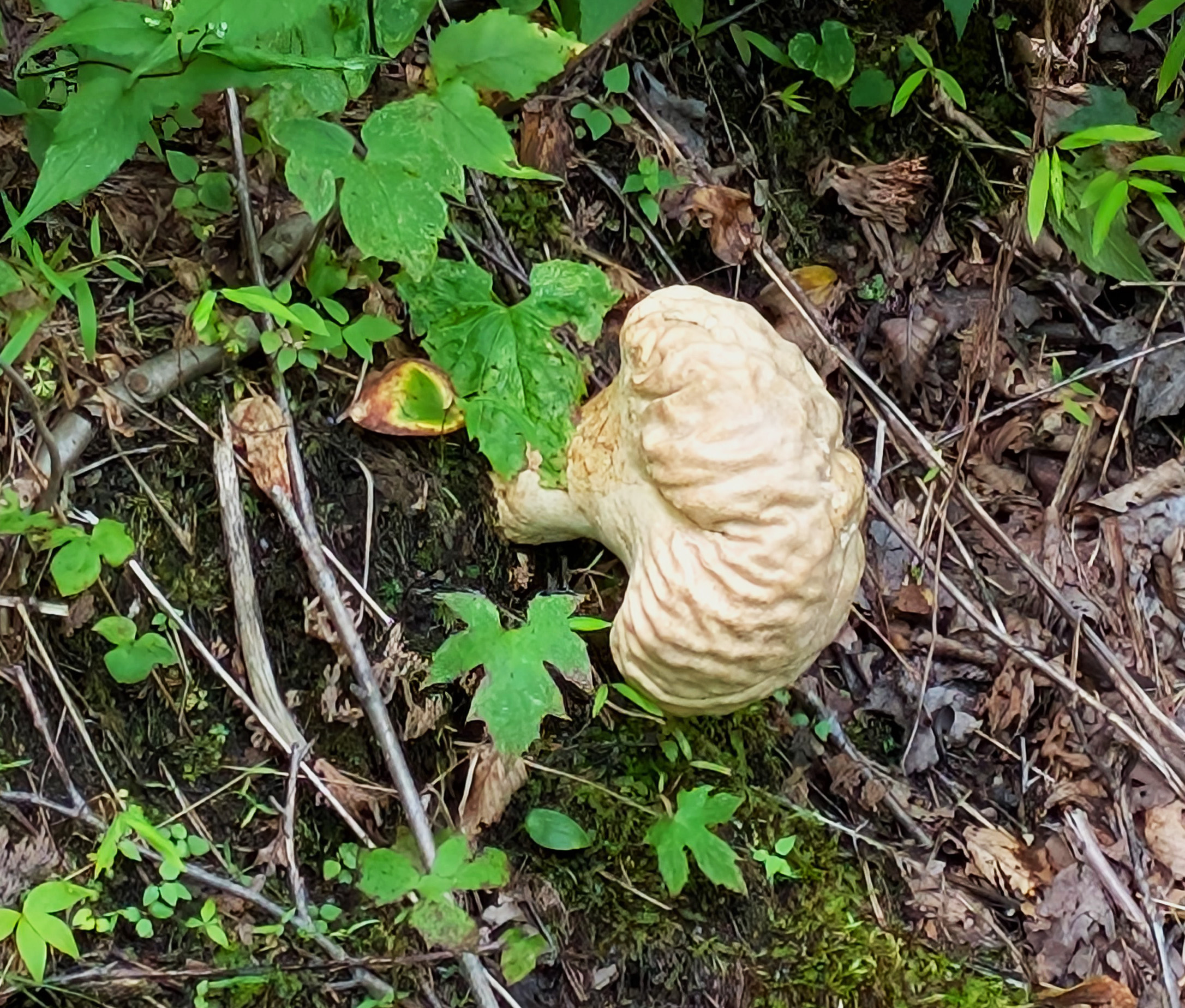
Calvatia craniiformis
Taxonomy
Domain: Eukaryota
Kingdom: Fungi
Division: Basidiomycota
Class: Agaricomycetes
Order: Agaricales
Family: Agaricaceae
Genus: Calvatia
Species: Calvatia craniiformis (Schwein.) Fr. ex De Toni (1888)
Synonyms: Bovista craniiformis Schwein. (1832); Calvatia craniiformis (Schwein.) Fr. (1849); Calvatia craniformis (orthographic variant)
Type species of the genus — Calvatia was erected around this fungus, and every other member of the genus, including the giant puffball, is measured against it.
A note on family placement: sources differ between Agaricaceae and Lycoperdaceae. The puffballs were formerly held in the order Lycoperdales, which molecular work dissolved; the reshuffling into Agaricales is settled, but the family assignment is still reported inconsistently across references.
Common Names by Region
United States / general: Brain puffball, skull-shaped puffball, brain-like puffball
Foraging usage: Skull puffball
Etymology: Calvatia from the Latin calvus, bald. Craniiformis is skull-shaped, from the same root as cranium. Both the scientific name and every common name are describing the same thing — the convoluted, folded surface of a mature fruit body, which looks unmistakably like a brain sitting on the forest floor.
Description
Fruit Body
- 8–20 cm broad by 6–20 cm tall. A large fungus.
- Ball-shaped when young, then developing a narrowed sterile base and taking on the form of an inverted pear or a skull
- White when young, aging through cream to tan and pale brown
- Attached to the substrate by a thick branching rhizomorph
Peridium (the skin)
- Smooth at first. Young specimens look like any other white puffball and are difficult to identify.
- Develops indentations, grooves, wrinkles, and folds as it matures — this is the diagnostic feature and the source of every name the fungus has
- Cracks and flakes with age, eventually sloughing away entirely to release the spore mass
Gleba (the spore mass)
- White and firm when young — uniform, structureless, and dense, with a texture often compared to tofu or fresh bread
- Matures through yellow to yellow-brown, greenish-yellow, and olive-brown, becoming powdery
- The color change is the single most important thing about this fungus from a practical standpoint, and it is discussed below
Sterile Base
- A persistent sterile base remains after the spores have gone
- The dried remnant can stand in the woods for a long time — these fruit bodies persist well after they have finished
Microscopic
- Spores globose to subglobose, 2.5–3.5 µm, nearly smooth
- Capillitial threads olive-colored, 2–4 µm wide, without sinuous slits
Quick ID
Large, tan, brain-folded mass with a narrowed base, sitting on the ground in open woods. Nothing else in eastern North America looks quite like a mature one.
Note on this specimen: at roughly the size of a child’s head, this is at the upper end of the recorded size range. The color and the fully developed brain folds indicate a mature fruit body — well past the white-gleba stage, and past edibility. This is a photographic specimen rather than a table one.
Known Range
North America — most common in the northeastern and southeastern United States, rarely found west of the Rocky Mountains. Also Asia and Australia. First reported from northeast India in 2018, and the distribution is probably still being filled in.
Habitat is open woods, grassy areas, wood edges, and roadsides.
Habitat & Ecology
- Saprobic — it decomposes organic matter in the soil and leaf litter rather than parasitizing anything.
- One 1966 study reported an ectomycorrhizal association with American sweetgum (Liquidambar styraciflua) under laboratory conditions, which sits oddly with the saprobic classification and has not been much followed up. Worth flagging as unresolved rather than settled.
- Fruits in summer and fall following rain, and in warm regions after any sustained wet period.
- Fruit bodies arise from the tips or lateral branches of rhizomorphs — the visible mushroom is one temporary structure on a much larger, mostly invisible organism running through the soil.
Look-Alikes and the Cut-Open Check
Field note — read this section before eating any puffball
The rule: cut every puffball in half from top to bottom, every time, before it goes anywhere near a pan.
A true puffball has a uniform white interior with no internal structure whatsoever. No cap outline, no gill lines, no stem, nothing.
An immature Amanita in the “egg” stage looks like a small white puffball from the outside. Cut in half, it reveals the developing cap, gills, and stipe in cross-section. Some Amanita species are lethal. This is the whole reason the check exists, it takes two seconds, and there is no acceptable substitute for it.
Other look-alikes worth knowing:
- Calvatia cyathiformis — similar size and habitat; the mature gleba turns purple-brown rather than olive.
- Handkea utriformis — externally similar, but develops a crater-like opening and has sinuous slits in the capillitial threads that C. craniiformis lacks.
- Calvatia gigantea, the giant puffball — much larger, staying smooth and white without the brain folds, and lacking the pronounced sterile base.
- Earthballs (Scleroderma) — thick rind, and the interior darkens to purple-black very early. Not edible.
Edibility
Edible only when the gleba is pure white, firm, and completely uniform. No intrinsic toxic compounds have been identified in the species, and it is widely eaten across North America, Asia, and Australia.
The window is narrow:
- Pure white throughout — good
- Any yellow coloration — the flavor turns bitter, and this is the signal to stop
- Yellow-brown or olive and powdery — this is spore mass. Not food.
- Any internal structure at all — not a puffball. Stop.
Texture in the good stage is firm and spongy, and the flavor mild. It takes on whatever it is cooked with, which is the usual reason people cook puffballs.
The standard cautions apply. Never eat a wild mushroom on the strength of a photograph or a website, including this one. Get it confirmed by someone who knows the local fungi, and try a small amount the first time — individual reactions to otherwise edible mushrooms are a real thing.
Traditional Medicine
Mature specimens have been used as a hemostatic and wound dressing in the traditional medicines of China and Japan, and by the Ojibwe. The dried spore mass is fine, absorbent, and was packed onto bleeding wounds — a use documented across several unconnected cultures on two continents, which is itself worth noting.
Modern laboratory work has found antibacterial and antifungal activity in extracts, including in a 2007 survey of nine Korean wild mushrooms. This is preliminary in vitro work and does not amount to a medical claim.
Additional Notes
- The spore output of puffballs is difficult to overstate. A single mature fruit body releases spores in numbers that run to the billions or beyond, dispersed by wind and by physical disturbance — rain, a falling branch, a passing animal, or a boot. The puff is the point.
- Two entirely different-looking fungi. A young C. craniiformis is a smooth white ball indistinguishable from a dozen other puffballs. A mature one is a tan folded brain. The same organism, weeks apart, and most identification confusion in this genus comes from that.
- The fruit body is the small part. What is visible is a temporary reproductive structure. The organism itself is a network of hyphae running through soil and litter, and it may have been there for years before producing anything above ground.
- It photographs well and it lasts. Because the sterile base persists, a fruit body found in one spot can often be relocated weeks later — useful for anyone documenting a site across a season.
Field Notes
- Look after rain, summer through fall, in open woods and along wood edges rather than in deep shade.
- Look on the ground. This is not a wood-decay fungus and will not be on a log.
- Cut it open whether or not you intend to eat it. The cross-section is the identification, and it teaches the eye faster than anything else.
- Leave mature ones standing. They are doing their job, they hold up well for photography, and they will disperse spores when something disturbs them.
- Photograph the base. The sterile base and the narrowed attachment are diagnostic and are the first thing lost when a specimen is picked up.
This site is open for educational use. Teachers and students are welcome to adapt, translate, and reuse this material with attribution.
Post Views: 84